NEXT STORY
The main ingredients in modern electron microscopy
RELATED STORIES
Video URL


NEXT STORY
The main ingredients in modern electron microscopy
RELATED STORIES


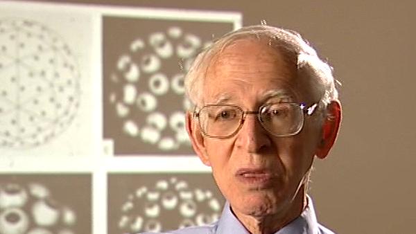
The image that was being produced in electron microscopy was mostly by what's called amplitude scattering, that is by scattering of the atoms of the molecule... of the object, scatter the electrons. But the... it was always an el... but we'd heard from Hugh Huxley, who was the... as I said the leading electron microscopist. Well, that to get a better picture, to get a better picture you should de-focus a bit and that seemed very odd to de-focus a bit. And it would be what's called under focussing rather that over focussing. And this seemed a bit of a mystery. And I... I knew the theory and I could work out the theory, the theory had been worked out by lots of people, the very complicated electron scattering theory. But it wasn't that that led to it, it was the... I decided that what we should do is to try to work out the difference between how much was amplitude contrast and how much was phase contrast. Phase contrast depends upon simply a shift in the position of the diffracted wave relative to some standard which you don't get in... in some scattering events. It's really too complicated to explain, but... so people... now, I wondered at this time how phase contrast arose, [Frits] Zernike in 1934 had given... was given the Nobel Prize for inventing the phase contrast micro... microscope. Now, that... that was the... what he did was to put a what's called a quarter way filter in the path of the... incident beam, which changes the phase relationship between the incident beam, which gets diffracted, and the diffracted rays. And this leads you to be able to see transparent objects. And I... I knew this, I began to wonder how it was that people could use the microscope to see transparent objects in the 1890s, 1900. And it turned out that old microscopes, you could see much better these, you could see... you could see transparent objects or detail very much better than you could in the more advanced microscopes, which corrected all the aberrations, the so called famous Cooke triplet. I read quite a lot about optics, history optics and realised that what was happening was that there was a mixture of phase contrast and amplitude contrast. And Harold Erikson was a Post Doc who came to the lab who was an expert electron microscopist, and we decided to study catalase, which is a simple two-dimension crystal to see if we could disentangle these two. And he did a series, a through-focal series on catalase and we discovered, I can't go into the whole details, the relative contribution of amplitude contrast and phase contrast. And the... it turned out that the, that Hugh Huxley's... Hugh Huxley's recipe for getting the best pictures was that he focussed by a very little bit. And John Finch discovered that... that a little bit had to be what's called 5,000 angstrom defocussing. If you defocus by anything more you got very, so called, sharp pictures but they were false detail. The pictures of Robert Horn, as one of the leading lights, turned out to be... looked very striking but they were... it was full of false detail; we analysed this by optical diffraction, in fact. So... so Harold's work led us to believe that you could recreate an image ... a transparent object by a series of taking defocussed pictures. You see, theoretically, a perfect phase object, perfect transparent object you can't image at all. However, if you actually defocus, any kind of variations then the then the density of the transparent material begin to show up. And so that at least you can get a picture, and by this time we'd introduced computer processing of images which replaced optical processing, which I'd introduced, and you could then correct the defocussed image in such a way to retrieve the original variation in density, giving rise to the phase variations. And so we suggested this, and it was the beginning of phase contrast and defocussing which is now in totally common use throughout. There are other things as well in electron microscopy, which is secondary scattering and so on but this was the biggest factor.
Born in Lithuania, Aaron Klug (1926-2018) was a British chemist and biophysicist. He was awarded the Nobel Prize in Chemistry in 1982 for developments in electron microscopy and his work on complexes of nucleic acids and proteins. He studied crystallography at the University of Cape Town before moving to England, completing his doctorate in 1953 at Trinity College, Cambridge. In 1981, he was awarded the Louisa Gross Horwitz Prize from Columbia University. His long and influential career led to a knighthood in 1988. He was also elected President of the Royal Society, and served there from 1995-2000.
Title: Electron microscopy: defocussing to see transparent objects
Listeners: Ken Holmes John Finch
Kenneth Holmes was born in London in 1934 and attended schools in Chiswick. He obtained his BA at St Johns College, Cambridge. He obtained his PhD at Birkbeck College, London working on the structure of tobacco mosaic virus with Rosalind Franklin and Aaron Klug. After a post-doc at Childrens' Hospital, Boston, where he started to work on muscle structure, he joined to the newly opened Laboratory of Molecular Biology in Cambridge where he stayed for six years. He worked with Aaron Klug on virus structure and with Hugh Huxley on muscle. He then moved to Heidelberg to open the Department of Biophysics at the Max Planck Institute for Medical Research where he remained as director until his retirement. During this time he completed the structure of tobacco mosaic virus and solved the structures of a number of protein molecules including the structure of the muscle protein actin and the actin filament. Recently he has worked on the molecular mechanism of muscle contraction. He also initiated the use of synchrotron radiation as a source for X-ray diffraction and founded the EMBL outstation at DESY Hamburg. He was elected to the Royal Society in 1981 and is a member of a number of scientific academies.
John Finch is a retired member of staff of the Medical Research Council Laboratory of Molecular Biology in Cambridge, UK. He began research as a PhD student of Rosalind Franklin's at Birkbeck College, London in 1955 studying the structure of small viruses by x-ray diffraction. He came to Cambridge as part of Aaron Klug's team in 1962 and has continued with the structural study of viruses and other nucleoproteins such as chromatin, using both x-rays and electron microscopy.
Tags: Hugh Huxley, Frits Zernike, Harold Erikson, John Finch, Robert Horn
Duration: 4 minutes, 59 seconds
Date story recorded: July 2005
Date story went live: 24 January 2008
