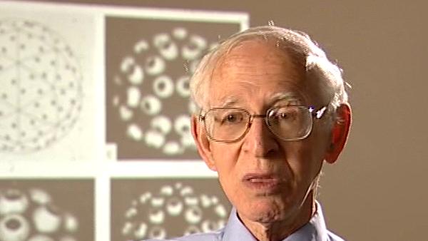
Once we developed three dimensional image reconstruction there was a clear parallel with an X-ray, X-ray photograph.
Yes.
And, so, I-
An ordinary medical photograph-
Ordinary medical photography, medical X-ray, medical X-radiology, but, you see, what they do is they take an X-ray picture and there are two ways of doing it. One is simply, you just, put up an X-ray film behind you, taken an X-ray picture and you get a picture of everything superimposed in the- in line. So, I- the other way was, a more sophisticated way, was called X-ray tomography. If you wanted a better view you had the- the, say the chest specimen, you wanted to see the different sections in greater detail. So, they had- so, what they do in medical tomography X-ray, tomography, was to have a moving source and a moving plate behind the back. And, the two things are moved at such a relative speed that only one plane in the line of sight is - not in focus, that's the wrong word, but is, you can see without blurring. All the rest is blown out-
Yes.
And, that's called X-ray tomography, which is not, which is used for special cases. The ordinary so-called X-rays just everything in the line of view. So, I- I did speak to a professor of radiology at Middlesex Hospital, a man called Windeyer, I knew- I got to know him because I knew his- one of his people in his department, an old South African friend of ours, so he told me about it. And, he said- oh, we don't need any of this fancy stuff or words to that effect. We know exactly what we're looking at, we don't need any of this. In any case, he said, if you're going to tilt the specimens, I'm not sure if he said it or one of the other consultants I spoke to. And, Uli Arndt, in fact, at Cambridge went to see the radiologist at Addenbrooks Hospital and he came with the same thing, we don't need any of this fancy stuff, we know exactly what we're looking at; which is true. They know the anatomy of the body and they know how to interpret the many- 1897 was the first X-ray, 60 years, 70 years of experience. So, and, what they said to me was, anyway if you're going to tilt the specimen you're going to take a lot of X-ray pictures and, the- you're going to expose the body to a lot of X-ray radiation and that can be very harmful. I was able to answer that, because I said, well, when you do an X-ray tomogram you're getting a lot of blurred detail and that means you're also exposing the specimen to unnecessarily- unnecessary radiation. But, I couldn't calculate at the time, somebody did that later, about the information gain you might get; it was quite a clever theoretical paper later on by- by having a series of tilts. But, what was unexpected was the fact that the- anyway, let me finish, but, still somebody read up, a man read up, it was a man called Godfrey Hounsfield, who was working for EMI. My paper with David Rowley was published in January 1968, in August 1968 Hounsfield took out a patent. He worked at EMI, Electronic Musical Instruments, but they also had a wide range of- they build electron microscopes for example. And, he took out at patent to build a device to collect data from 60, it had 60 detectors and you, you, the idea was you would take a series of X-ray pictures and then you would assemble them by three dimensional image reconstruction. Now, what he patented was not the idea, he patented the instrument. Now, the idea he told me that he'd read all our papers, one, only two at the time, he'd read the papers and he'd followed them. And, in fact, in 1975 we had an exhibition at the Royal Society, a soiree, which Tony Crowther and I and I think you as well, John, I might remember, illustrated three dimensional image reconstruction in electron microscopy. And, he illustrated three dimensional image reconstruction in medical radiography. So, he was the- he got the Nobel Prize together with a man called Allan Cormack in 1979. I was a bit put out by it because I don't think there's any doubt that he followed what I did, in fact, he more than followed what I did because when he, well, some people think I should have shared it but it didn't matter in the end because I got a Nobel Prize three years later in Chemistry
Born in Lithuania, Aaron Klug (1926-2018) was a British chemist and biophysicist. He was awarded the Nobel Prize in Chemistry in 1982 for developments in electron microscopy and his work on complexes of nucleic acids and proteins. He studied crystallography at the University of Cape Town before moving to England, completing his doctorate in 1953 at Trinity College, Cambridge. In 1981, he was awarded the Louisa Gross Horwitz Prize from Columbia University. His long and influential career led to a knighthood in 1988. He was also elected President of the Royal Society, and served there from 1995-2000.
Title: 3D imaging: X-ray tomography and Godfrey Hounsfield's patent
Listeners:
John Finch
Ken Holmes
John Finch is a retired member of staff of the Medical Research Council Laboratory of Molecular Biology in Cambridge, UK. He began research as a PhD student of Rosalind Franklin's at Birkbeck College, London in 1955 studying the structure of small viruses by x-ray diffraction. He came to Cambridge as part of Aaron Klug's team in 1962 and has continued with the structural study of viruses and other nucleoproteins such as chromatin, using both x-rays and electron microscopy.
Kenneth Holmes was born in London in 1934 and attended schools in Chiswick. He obtained his BA at St Johns College, Cambridge. He obtained his PhD at Birkbeck College, London working on the structure of tobacco mosaic virus with Rosalind Franklin and Aaron Klug. After a post-doc at Childrens' Hospital, Boston, where he started to work on muscle structure, he joined to the newly opened Laboratory of Molecular Biology in Cambridge where he stayed for six years. He worked with Aaron Klug on virus structure and with Hugh Huxley on muscle. He then moved to Heidelberg to open the Department of Biophysics at the Max Planck Institute for Medical Research where he remained as director until his retirement. During this time he completed the structure of tobacco mosaic virus and solved the structures of a number of protein molecules including the structure of the muscle protein actin and the actin filament. Recently he has worked on the molecular mechanism of muscle contraction. He also initiated the use of synchrotron radiation as a source for X-ray diffraction and founded the EMBL outstation at DESY Hamburg. He was elected to the Royal Society in 1981 and is a member of a number of scientific academies.
Tags:
Middlesex Hospital, Addenbrooks Hospital, Godfrey Hounsfield, Uli Arndt, Anthony Crowther, Allan Cormack
Duration:
5 minutes, 15 seconds
Date story recorded:
July 2005
Date story went live:
24 January 2008


